Human cheek cell lab report introduction Image result for human cheek cell diagram Cheek cells under a microscope
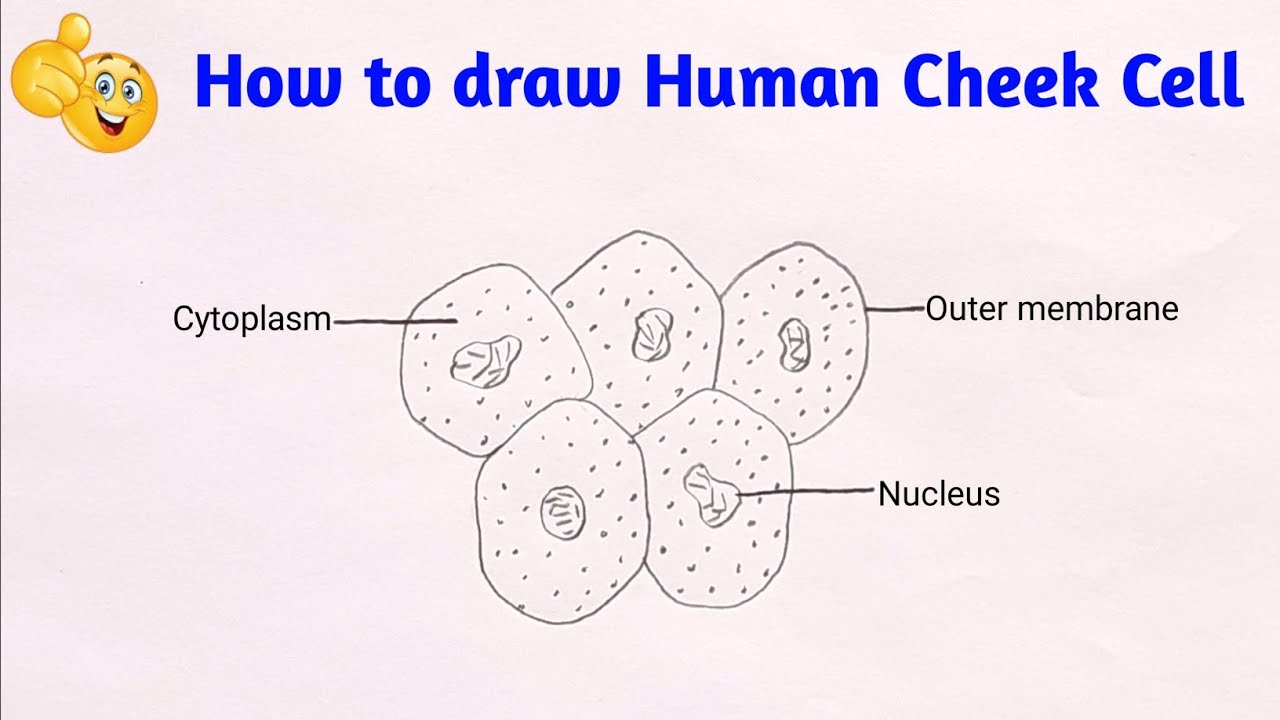
Cheek Cell Labeled Diagram
Cheek cell diagram
Cheek cells conclusion
Solved using this table from the size estimation module,Solved using this table from the size estimation module, Cheek cell size cells human using 40x objective single module estimation table lens field organelle well solved determine writeDraw the diagram of cheek cells and label the parts..
Cell cheek cells 400x stained human animal slide lab staticflickr picture c1 flickrCheek cell image using brightfield and darkfield microscopy. (a Cbse class 9 science practical skills – slide of onion peel and cheek cellsHuman epidermal cells diagram.

Cheek cell image using brightfield and darkfield microscopy. (a
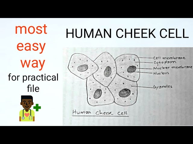
Diagram of cheek cellsDraw the human cheek cell with correct labelling How to draw cheek cell step by step for beginnersDraw the picture of cheek cells andlare ita.
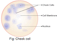
Cheek cells under microscope labeledCheek cell bacteria cells human membrane nucleus using picture bacterial been single prokaryotic solved determine Draw cheek cellCheek onion cell vs cells comparing contrasting.

Cheek brightfield
Cheek cells under microscope labeledSbi3u Cheek cell diagramCheek labelling ppz brainliest.
How would you take the sample to prepare temporary stained mount ofDarkfield microscopy brightfield cheek condenser Human cheek cells under the microscopeDraw three types of cells (cheek cell, red blood cell, elodea). make.

5. describe an activity to observe cheek cells under a microscope.
Drawing cell cheek labelled human biological parts followed rules basic must there when some exportedTop 197 + animal cheek cell Cheek cell human stained temporary cells mounts prepare epithelial lab results layer work discussion studyDraw the human cheek cell with correct labelling.
Cheek cell labeled diagramSquamous epithelial cheek cells labeled Cheek cell human draw labelling correctCheek cells 400x stained.

Dic image of a cheek cell
Human cheek cell dna extractionCheek dna extraction chromosomes mugeek vidalondon genetic Cheek labeled membrane nucleus elodea drawings.
.